3.6 Connections between Cells
Introduction
You already know that tissue is a group of similar cells working together. As you might expect, if cells are to work together, they must communicate with each other, just as you need to with others if you work on a group project. Consider how cells interact with their environment and communicate with each other. Cell-surface proteins govern interactions between a cell and its environment or with other cells. This Topic examines a subset of those interactions: direct cell contact with either other cells or the extracellular matrix (ECM).
The ECM is a general term for the extremely large proteins and polysaccharides secreted by some cells in a multicellular organism. They act as connective material to hold cells in a defined space. Cell density can vary greatly between different tissues of an animal, from tightly packed muscle cells with many direct cell-to-cell contacts to liver tissue, in which some of the cells are only loosely organized and suspended in a web of extracellular matrix.
Unit 3, Topic 6 Learning Objectives
By the end of Unit 3, Topic 6, you will be able to:
- Describe the components of the ECM.
- Describe how the ECM and cytoskeleton interact.
- List examples of the ways that plant cells and animal cells communicate with adjacent cells.
- Summarize the roles of tight junctions, desmosomes, gap junctions, and plasmodesmata.
| Unit 3, Topic 6—To Do List | Suggested Average Time to Complete (min.) |
|---|---|
| Read, make summary notes, and complete the self-check questions for Unit 3, Topic 6 of TRU Cell and Molecular Biology. | 60 |
| Complete Learning Activity: Cell-extracellular matrix mechanobiology. | 10 |
| Complete Learning Activity: Extracellular matrix. | 10 |
| Complete Learning Activity: Extracellular matrix—Khan Academy. | 15 |
| Complete Learning Activity: Integrins: The receptors that keep it together. | 6 |
| Complete Learning Activity: Cell junctions. | 10 |
| Go to Moodle and complete the Unit 3 Quiz. | 7 |
| Go to Moodle and complete the Unit 3 Assignment. | 100 |
The Extracellular Matrix of Animal Cells
While cells in most multicellular organisms release materials into the extracellular space, animal cells specifically will be discussed here. The extracellular matrix (ECM) consists of proteins and polysaccharides secreted by cells and assembled into an organised meshwork external to the cell. ECM is a generic term encompassing mixtures of polysaccharides and proteins, including collagens, fibronectins, laminins, and proteoglycans, all secreted by the cell (Figure 1). The primary components of the ECM are proteins, and the most abundant protein is collagen. Collagen fibres interweave with proteoglycans, which are carbohydrate-containing protein molecules.
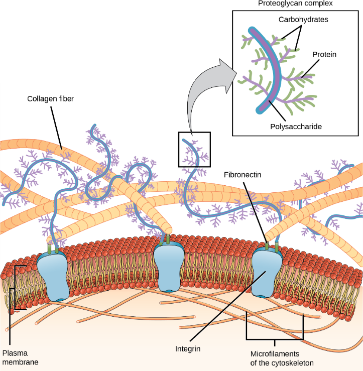
The amount and type of ECM varies from tissue to tissue, and different types of ECM perform specific functions. ECM comes in two relatively different examples. First is the basement membrane underlying the epidermis of the skin. The basement membrane is a thin, almost two-dimensional layer that helps organize skin cells into a nearly impenetrable barrier to most simple biological insults. Second is the massive three-dimensional matrix surrounding each chondrocyte in cartilaginous tissue. The ability of the cartilage in a person’s knee to withstand the repeated shock of their footsteps is due to the ECM proteins in which the cells are embedded, not to the cells that are relatively few and sparsely distributed. Although both ECM types share some components in common, they are clearly distinguishable in their function, appearance, proportions, and identity of the constituent molecules. The extracellular matrix holds the cells together to form tissues and allows the cells within the tissue to communicate with each other. The ECM also serves as a reservoir for extracellular signalling molecules that control cell growth, migration, and differentiation.
Blood clotting is an example of the extracellular matrix’s role in cell communication. When the cells lining a blood vessel are damaged, they display a protein receptor called tissue factor. When tissue factor binds with another factor in the extracellular matrix, it causes platelets to adhere to the damaged blood vessel’s wall, stimulates the adjacent smooth muscle cells in the blood vessel to contract (thus constricting the blood vessel), and initiates a series of steps that stimulate the platelets to produce clotting factors.
The Difference Between Basal Lamina and Basement Membrane
Basal lamina is a specialized form of extracellular matrix underlying the basal side of polarized epithelial cell sheets that separates them from the underlying connective tissue.1 The “basal lamina” and “basement membrane” are frequently confused by students and professionals alike. They found a very thin layer of connective proteins just beneath an epithelial cell layer. Meanwhile, the basal lamina was not discovered until later because it is not visible by light microscopy (usually only ~50 nm thick). Technically, the basal lamina, which consists of multiple layers, is a layer of ECM proteins secreted by the epithelial layer. The basal lamina and a thick reticular lamina (ECM secreted by other cell types) form the basement membrane.
Self-Check
Explain how the extracellular matrix functions in your own words using terminology in the Unit 3, Topic 6 readings.
Show/Hide answer.
The extracellular matrix acts as a support and attachment in animal tissues. It holds cells together and facilitates cell communication. It also functions in the healing and growth of the tissue and blood clotting.
In most animal tissues, the extracellular matrix is secreted mainly by cells called fibroblasts, and the main component is a gel-like substance with embedded proteins. The gel is composed of polysaccharides called glycosaminoglycans (GAGs), which are often attached to proteins to form heavily glycosylated proteins called proteoglycans. The GAGs are negatively charged and attract positively charged ions, particularly sodium ions. The high positive ion concentration leads to water moving via diffusion into the matrix, which gives the matrix its gel-like properties, including the ability to resist compression.
Three major types of macromolecules make up the extracellular matrix:
- Structural proteins
- Adhesive proteins
- Proteoglycans
Structural Proteins
The two matrix proteins are collagen and elastin, both fibrous proteins.
Collagen is the main protein in the extracellular matrix. It accounts for 25% of the total protein content in a human organism. Collagen consists of three polypeptide chains wound around one another to form a triple helical structure. Gly-X-Y is the primary amino acid sequence, whereby X is often proline.
There are at least 30 different collagen peptide chains. Types I-IV are of primary significance. The most abundant type is collagen type I, one of the fibril-forming collagens that are the basic structural components of connective tissues. The principal function of collagen is to strengthen and organize the ECM.
Elastin is another matrix protein that is the main component of elastic fibres. An elastin molecule consists of flexible polypeptide chains and can reversibly unroll under mechanical stretching forces. These fibres provide elasticity to blood vessels, the lungs, skin, and the ligaments.
Adhesive Proteins
Matrix adhesive proteins, like fibronectin and laminin, mediate the connection between the components of the matrix to one another and to the surfaces of cells. Fibronectin is one of the adhesive glycoproteins providing the attachment of cells to the extracellular matrix. Laminins mediate the attachment of parenchymal cells to type IV collagen, thereby providing the interaction between cells and basement membranes. Other extracellular matrix glycoproteins are nidogens, tenascins, and fibulins.
Proteoglycans
Proteoglycans are composed of proteins covalently attached to long nonbranched chains of polysaccharides, glycosaminoglycans.
The polysaccharide content is more than 95% in the proteoglycans. These polysaccharides are highly negatively charged and bind positively charged ions and water molecules to form hydrated gels; this characteristic controls how small molecules diffuse and resist compressive forces. Glycosaminoglycans occupy large volumes in tissues, forming strongly hydrated gels that cause tissue turgor. The turgor gives the tissue the ability to resist compression forces. For example, an articular cartilage can resist mechanical pressures of a hundred atmospheres.
Proteoglycans can form huge polymeric complexes in the extracellular matrix. Besides providing tissue turgor, proteoglycans can also connect to other extracellular matrix proteins, forming complex structures (e.g., basement membranes).
Learning Activity: Cell-extracellular Matrix Mechanobiology
- Watch the video “Cell-Extracellular Matrix Mechanobiology” (3:48 min) by SciTube (2019).
- Explain in your own words what the following paragraph is describing after seeing this animation:
“Cells have protein receptors on their plasma membranes’ extracellular surfaces. When a molecule within the matrix binds to the receptor, it changes the receptor’s molecular structure. The receptor, in turn, changes the microfilaments’ conformation positioned just inside the plasma membrane. These conformational changes induce chemical signals inside the cell that reach the nucleus and turn “on” or “off” the transcription of specific DNA sections, which affects the associated protein production, thus changing the activities within the cell. Watch this scenario unfold in the following video.”
Learning Activity: Extracellular Matrix
- Watch the video “Extracellular matrix” (4:16 min) by Kathy Papastephanou (2017).
- Answer the following questions based on your Unit 3, Topic 6 readings and information provided in the video:
- Why does a multicellular environment require an ECM? When during human life does multicellular communication begin?
- What is the name of the system that connects cells and allows them to communicate?
- What three components comprise the ECM? Where is it relative to the cell?
- How is the ECM connected to the cytoskeleton of cells?
- What are the three roles of the ECM mentioned in the video?
Learning Activity: Extracellular Matrix — Khan Academy
- Watch the video “Extracellular matrix | Structure of a cell | Biology | Khan Academy” (6:53 min) by Khan Academy (2015).
- Answer the following questions based on your Unit 3, Topic 6 readings and information provided in the video:
- What is the most abundant protein in mammals?
- What transmembrane protein do cytoskeletal actin microfilaments connect to?
- To which transmembrane protein do ECM proteins collagen and fibronectin connect?
- Draw this inside-outside connection yourself.
- Is everything known about these connections?
- Go to the National Library of Medicine, National Center for Biotechnology Information ([date unknown]) website and find a recent primary research article (2021 and up) on one or more of the components in your drawing.
Learning Activity: Integrins — The Receptors That Keep It Together
- Watch the video “Integrins: The receptors that keep it together” (3:01 min) by the Albert and Mary Lasker Foundation (2022).
- Answer the following questions based on the information provided in this video:
- What did researchers R. Hynes and E. Ruoslahti find when comparing proteins in healthy and cancer cells in the 1970s? What did they start looking for?
- What was researcher Timothy Springer looking for?
- What did Ruoslahti discover 10 years later in the 1980s? What did this finding lead to?
- How many of these newly discovered proteins have been now found in mammals?
Intercellular Junctions
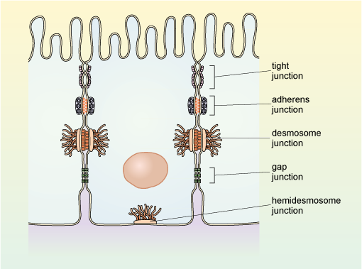
Cells can also communicate with each other via direct contact or intercellular junctions. There are differences in how plant, animal, and fungal cells communicate. Plasmodesmata are junctions between plant cells; whereas, animal cell contacts include tight junctions, gap junctions, and desmosomes (Figure 2).
Epithelial tissue, composed of closely packed sheets of cells, has several types of cell junctions, each with specific functions and formed by different specialized proteins. In vertebrates, there are the following three major types of cell junctions:
- Tight junctions (occluding junctions)
- Adherens junctions, desmosomes and hemidesmosomes (anchoring junctions)
- Gap junctions (communicating junctions)
Plasmodesmata
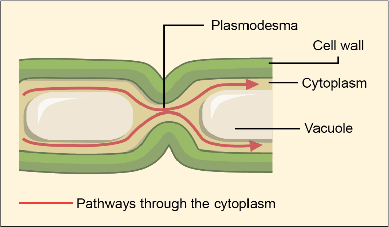
In general, long stretches of the plasma membranes of neighbouring plant cells cannot touch one another because the cell wall surrounding each cell separates them. Then, how can a plant transfer water and other soil nutrients from its roots, through its stems, and to its leaves? Such transport primarily uses the vascular tissues (xylem and phloem). Structural modifications, called plasmodesmata (singular plasmodesma), also exist. Numerous channels passing between adjacent plant cells’ cell walls connect their cytoplasm and enable the transport of materials from cell to cell and, thus, throughout the plant (Figure 3).
Tight Junctions
A tight junction is a watertight seal between two adjacent animal cells (Figure 4). Protein complexes tightly hold the cells against each other. The basic functional units forming adhesive contacts across cells are the tight junction strands, which are composed of transmembrane proteins such as claudin, occludin and tricullulin. Assembling these strands requires them to associate with a cytoplasmic plaque of adaptor proteins, such as the ZO proteins, which link them to several regulatory and signalling molecules and to the cortical cytoskeleton. Coordinated interactions amongst the molecular core regulate tight junction assembly by ensuring proper localization of tight junction components and stabilizing the junctional complex.
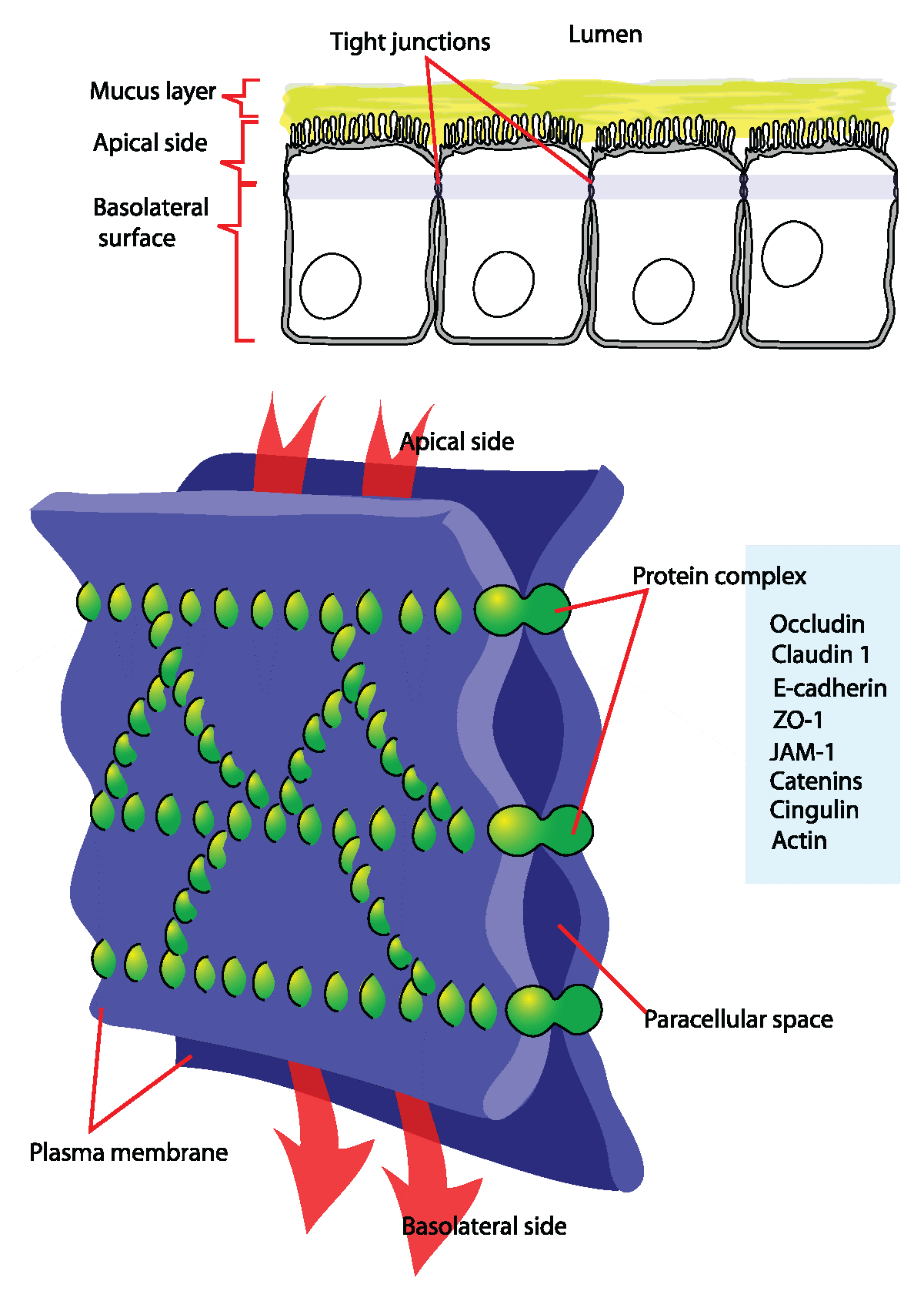
This tight adherence prevents materials from leaking between the cells; tight junctions are typically found in epithelial tissues lining internal organs and cavities and comprise most of the skin. For example, epithelial cells lining the urinary bladder have tight junctions to prevent urine from leaking into the extracellular space. In the digestive system, they help prevent digestive enzymes from leaking into the bloodstream. Tight junctions also serve as a structural support mechanism that helps keep the epithelium together.
Self-Check
Pathogenic E. coli have recently been shown to degrade tight junction proteins during infection. How would this provide an advantage to the bacteria?
Show/Hide answer.
E. coli infections generally cause food poisoning, meaning that the invading bacteria cross from the lumen of the gut into the rest of the body. Tight junctions hold the epithelial layer lining the digestive tract together so that the material that crosses into the body is tightly regulated. One way E. coli can avoid this regulation is to destroy the tight junctions so it can enter the body between the epithelial cells rather than going through the cells.
Adherens Junctions
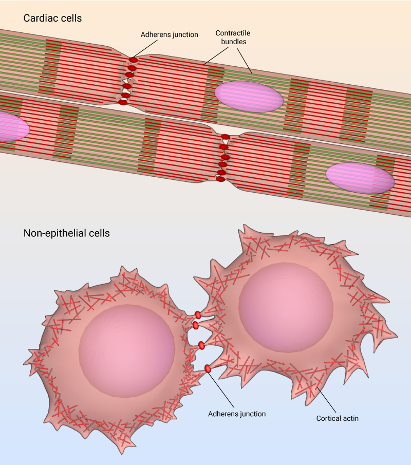
Adherens junctions are cell-cell adhesion complexes that the body continuously assembles and disassembles, allowing cells within tissue to respond to forces, biochemical signals and structural changes in their microenvironment. Adherens junctions are also called zonula adherens, intermediate junctions, or belt desmosomes. Zonula means small zone or belt-like, and adherens refers to adhesion (sticking together). As a result, the zonula adherens often run like a belt in a continuous fashion around the entire cell and act as an adhesion belt. It contains contractile bundles of actin filaments, usually located near the apical surface of polarized epithelial cells.
This type of cell junction is located right below tight junctions and provides a strong bond between the sides of adjacent epithelial cell membranes (Figure 2). While other junctions, like tight junctions, provide some support for and fusion of adjacent cells, their resistance to mechanical stress is relatively small compared to the much stronger adherens junctions. Adherens junctions come in many forms: In cardiac cells, adherens junctions anchor contractile bundles2, while in nonepithelial tissues, adherens junctions link cortical actin filaments between cells (Figure 5).
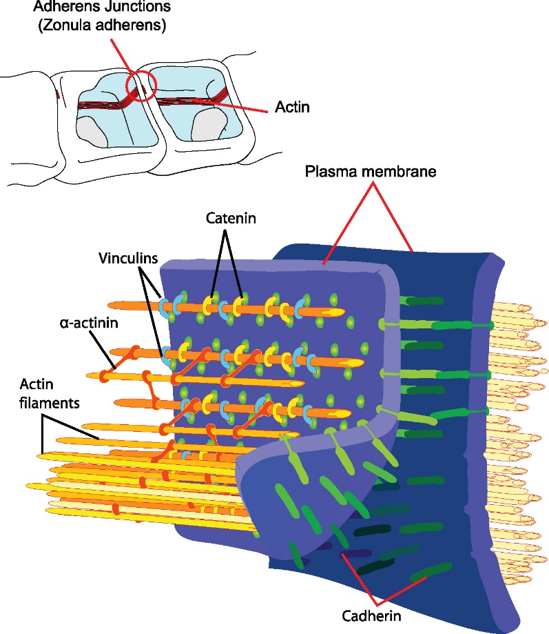
The zonula adherens is composed of the following different proteins (Figure 6):
- Actin microfilaments of the cytoskeleton (the internal skeleton of the cell)
- Cadherins, namely E-cadherin. These are transmembrane adhesion proteins, whose main portions are in the extracellular space.
- Anchor proteins are inside each cell. These are called α-catenin, β-catenin, γ-catenin (aka plakoglobin), vinculin, and α-actinin. They link the actin microfilaments to the cadherins.
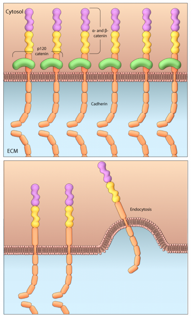
The extracellular part of one cell’s cadherin binds to the extracellular part of the adjacent cell’s cadherin in the space between the two cells. Each cell’s cadherin molecule also contains a tail that inserts itself inside its respective cell (Figure 7).
Mechanobiology Institute, National University of Singapore
This intracellular tail then links to catenin proteins to form the cadherin–catenin complex. Catenins are a family of proteins found in complexes with cadherin cell adhesion molecules of animal cells. The first two catenins identified3 became known as α-catenin and β-catenin. α-Catenin can bind to β-catenin and F-actin.4 β-Catenin binds directly to the cytoplasmic tail of classical cadherins (Figure 7). This complex binds to vinculin and α-actinin; these two proteins link the cadherin–catenin complex to the cell’s internal skeletal framework (the actin microfilaments).
Bonding the extracellular portions of the cadherin molecules in adjacent cells together requires calcium ions (or another protein in some cases). This means that the functional and morphological integrity of the adherens junctions are calcium-dependent. Removing calcium would result in this type of cell junction disintegrating.
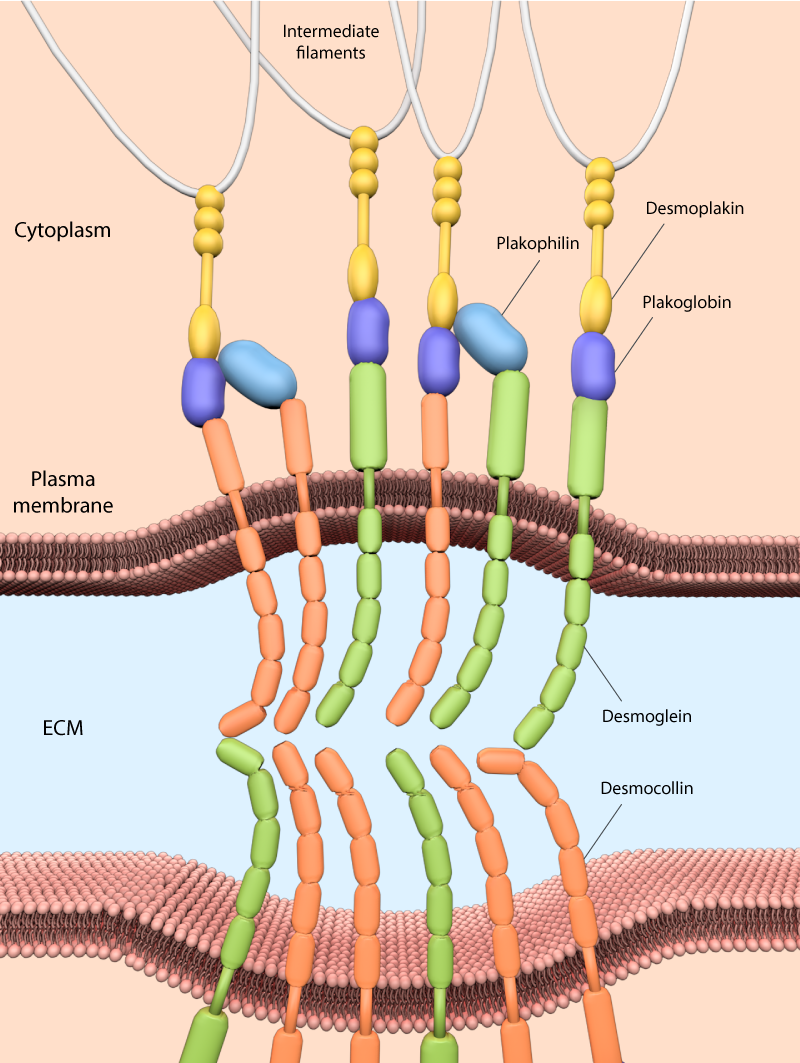
Desmosomes
Only animal cells have desmosomes, which act like spot welds between adjacent epithelial cells (Figure 8). These junctions link intermediate filaments to the plasma membrane and, in doing so, provide mechanical stability to cells, which is particularly important for tissues and organs under high mechanical stress (e.g., myocardium, skin, and bladder).
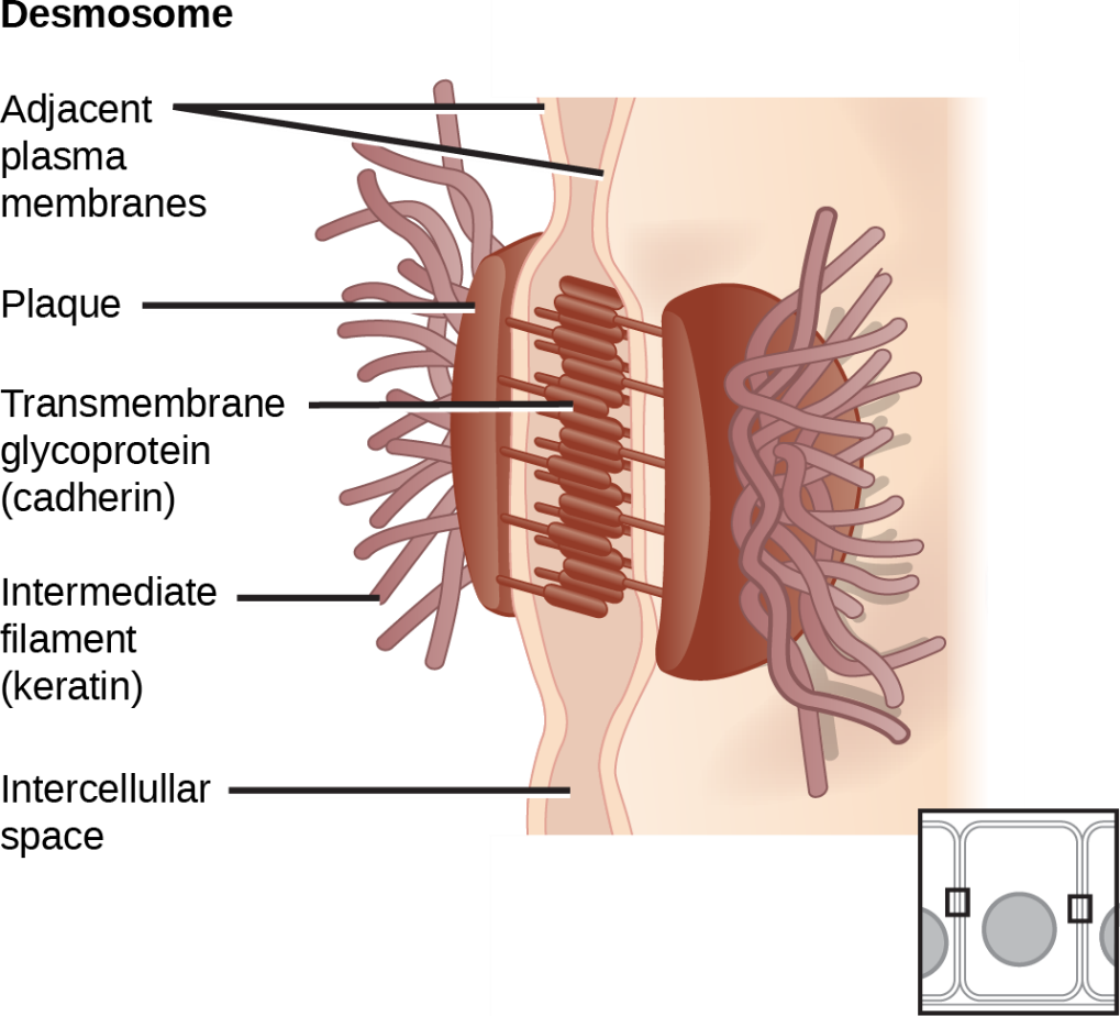
Desmosomes comprise desmosomal cadherins (desmogleins and desmocollins) that facilitate linkage between apposing cells via their extracellular domains and bind other desmosomal components intracellularly via their cytoplasmic tails (Figure 9). Cytoplasmic desmosomal components include plakoglobin and plakophilins, which bind intermediate filaments via desmoplakin. These cadherins connect two adjacent cells and maintain the cells in a sheet-like formation in organs and tissues that stretch, like the skin, heart, and muscles.
Mechanobiology Institute, National University of Singapore
Gap Junctions
Gap junctions in animal cells are like plasmodesmata in plant cells in that they are channels between adjacent cells that allow for transporting ions, nutrients, and other substances that enable cells to communicate. Structurally, however, gap junctions and plasmodesmata differ. Gap junctions are in many places throughout the body, including the epithelia (covering body surfaces), nerves, cardiac (heart) muscle, and smooth muscle (e.g., the intestines). Their primary role is to coordinate the activity of adjacent cells. For example, when heart cells need to beat in unison, gap junctions allow for the transmission of electrical signals between the cells.
Each gap junction channel has two half channels (hemichannels), one in each cell’s membrane (Figure 10). Each of these half channels is called a connexon. Each connexon has six symmetrical integral membrane protein units called connexins. This composition means each channel has 12 circularly arranged protein units. These half channels join together, bridge the extracellular space in the process, and form the entire channel that spans both cell membranes (Figure 11).
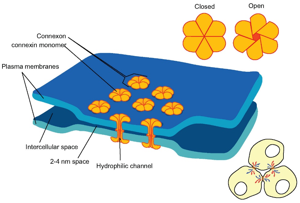
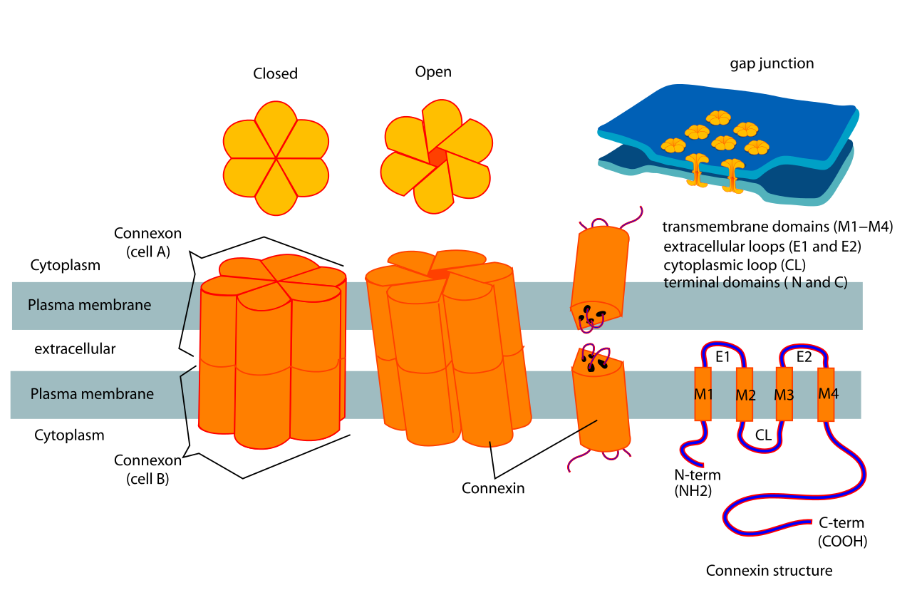
When the connexon’s pores (“doughnut holes”) in adjacent animal cells align, a channel between the two cells forms. Gap junctions are especially crucial in cardiac muscle. The electrical signal for the muscle to contract passes efficiently through gap junctions and allows the heart muscle cells to contract in tandem. The molecules that may cross this channel include ions, regulatory proteins, and metabolites (products of metabolism). Examples include calcium ions and cAMP (cyclic adenosine monophosphate).
Depending on the type of gap junction in question, molecules can pass evenly in both directions or asymmetrically, meaning the molecules move in one direction faster than in the other direction. The channels in a gap junction are not always open. They fluctuate between being open and closed. The ability of the channel to open or close is made possible in part due to calcium ions, which induce a reversible conformational change in the connexin molecules, leading to a channel closing at its extracellular surface. The cytoplasmic end of each connexon can also be closed if necessary.
Learning Activity: Cell Junctions
- Watch the video “Cell Junctions” (5:57 min) by Gaylene Ewing (2013).
- Answer the following questions based on the information provided in this video:
- What is a good way to remember the function of a tight junction? Where in the body are tight junctions found?
- What major function of adherens junctions did the video discuss?
- In desmosomes, cadherins attach to what cytoskeletal component?
- What function do desmosomes serve?
- What is the purpose of gap junctions?
- What is the name of the tunnel between cells in gap junctions? Use the terminology in Unit 3, Topic 6 to describe and draw the structure of these tunnels.
Key Concepts and Summary
The cells of animal tissues are held together by the extracellular matrix and by cell junctions.
The extracellular matrix in animals consists of a hydrated mixture of glycosaminoglycans (GAGs) and proteoglycans, in which various proteins, especially fibrous proteins like collagen, are embedded. The properties of the matrix, which vary from tissue to tissue, determine the nature of the protein.
Cells of epithelia are closely connected and not separated by intracellular material. Three basic types of connections allow varying degrees of interaction between the cells: tight junctions, anchoring junctions (adherens, desmosomes) and gap junctions.
- Tight junctions are found in epithelial and endothelial cells and primarily function as diffusion barriers.
- Adherens junctions regulate cell shape, maintain tissue integrity and translate actomyosin-generated forces throughout a tissue.
- A gap junction is a channel of two half channels called connexons that allow various molecules and ions to pass freely between cells. Gap junctions allow for electrical communication between cells, which is why they are most notable in neurons and cardiac cells.
Cell junctions form from specialized membrane proteins that connect to various cytoskeletal components such as actin filaments (adherens junctions) or intermediate filaments (desmosomes).
Various cadherin molecules comprise adherens junctions and desmosomes.
All three previously mentioned junctions in vertebrate epithelia have defined spatial organizations, with tight junctions located most apically, followed by adherens junctions and desmosomes.
Quiz
Complete the Unit 3 Quiz found in the Assessments Overview section in Moodle.
Assignment
Complete the Unit 3 Assignment found in the Assessments Overview section in Moodle.
Key Terms
adherens junctions
multi-molecular complexes beneath tight junctions that link the actin cytoskeleton to the plasma membrane via transmembrane proteins (cadherins) and intracellular proteins, thereby forming adhesive belts between cells
anchoring junctions
(includes adherens junctions, desmosomes and hemidesmosomes). Intercellular junctions that are present in many types of animal tissues and serve to hold the constituent cells to each other and to the surrounding extracellular matrix respectively, via transmembrane proteins, cadherins
basal lamina
a specialized layer of extracellular matrix lying basal to epithelial cells and separating them from the underlying connective tissue. The basal lamina is secreted by both epithelial cells and connective tissue cells and is primarily composed of glycoproteins and proteoglycans
cadherin
any of a class of transmembrane proteins important in maintaining tissue structure
catenin
a family of proteins found in complexes with cadherin cell adhesion molecules of animal cells; the first two catenins that were identified became known as α-catenin and β-catenin
cell adhesion molecule
help cells stick to each other and to their surroundings; the proteins located on the cell surface bind with other cells or with the extracellular matrix (ECM)
claudins
proteins that form the backbone of the tight junction strands
collagen
a protein that is abundant in the extracellular matrix. There are several types of collagen; it can form long thin fibres and provide structure, strength and flexibility to many tissues
desmosome
linkages between adjacent epithelial cells that form when cadherins in the plasma membrane attach to intermediate filaments
extracellular matrix (ECM)
non-cellular material secreted from animal or fungal cells that provides mechanical protection and anchoring for the cells in the tissue
gap junction
is a type of junction mediated by membrane channels called connexons that are used to transfer information between neighbouring cells (e.g., electrical signals, chemical messengers, etc.)
peptidoglycan
major component of bacterial cell walls. It is a polymer of high molecular mass, composed of two complex monosaccharides derived from glucose, which are linked together by amino acids, including three amino acids that are not found naturally in proteins
plasmodesma (plural plasmodesmata)
channel that passes between adjacent plant cells’ cell walls, connects their cytoplasm, and allows transporting of materials from cell to cell
tight junctions
a type of junction that forms tight contacts between cells in animal tissues such as epithelia. Tight junctions prevent the lateral movement of other membrane proteins, thus allowing different regions of the cell membrane to have different functional properties (e.g., transport characteristics)
Media Attributions
- Figure 1: Figure 4.27 from OpenStax Biology 2e (Clark et al. 2018) is used under a CC BY 4.0 license.
- Figure 2: Figure 26 from The Open University A Tour of the Cell (The Open University 2017) is used under a CC BY-NC-SA 4.0 license.
- Figure 3: Figure 4.28 from OpenStax Biology 2e (Clark et al. 2018) is used under a CC BY 4.0 license.
- Figure 4: Tight junction: Diagram of… from Anatomy and Physiology (Boundless) (Anatomy … 2023) is used under a CC BY-SA 4.0 license.
- Figure 5: Adherens junction in cardiac… by Mechanobiology Institute (2023) used with permission.
- Figure 6: The structural proteins in from Anatomy and Physiology (Boundless) (Anatomy … 2023) is used under a CC BY-SA 4.0 license.
- Figure 7: Scaffolding protein p120-catenin… by Mechanobiology Institute (2023) used with permission.
- Figure 8: Figure 4.29 [modification of work by Mariana Ruiz Villareal] from OpenStax Biology 2e (Clark et al. 2018) is used under a CC BY 4.0 license.
- Figure 9: Desmosome junctions link… by Mechanobiology Institute (2023) used with permission.
- Figure 10: Gap junction: The major… from Anatomy and Physiology (Boundless) (Anatomy … 2023) is used under a CC BY-SA 4.0 license.
- Figure 11: Connexon and connexin structure by Mariana Ruiz [LadyofHats] (2006), via Wikimedia Commons, is in the public domain.
References
A tour of the cell. 2017. Milton Keynes (England): The Open University; [accessed 2024 Jan 22]. Glossary. https://www.open.edu/openlearn/science-maths-technology/a-tour-the-cell/content-section–glossary.
Albert and Mary Lasker Foundation. Integrins: the receptors that keep it together [Video]. YouTube. 2022 Sep 28, 3:01 minutes. [accessed 2024 Jan 29]. https://www.youtube.com/watch?v=4k60P3Pnh30.
Anatomy and physiology (Boundless). 2023. Westchester (IL): Follett; [accessed 2024 Jan 18]. https://med.libretexts.org/Bookshelves/Anatomy_and_Physiology/Anatomy_and_Physiology_(Boundless).
Anatomy and physiology (Boundless). 2023. Westchester (IL): Follett; [accessed 2024 Jan 18]. Chapter 5.2A: tight junctions, Chapter 5.2B: adherens junctions, Chapter 5.3C: gap junctions. Figures Tight junction: diagram of…, Adherens junction in cardiac…, Gap junction: the major. https://med.libretexts.org/Bookshelves/Anatomy_and_Physiology/Anatomy_and_Physiology_(Boundless).
4 Buckley CD, Tan J, Anderson KL, Hanein D, Volkmann N, Weis WI, Nelson WJ, Dunn AR. 2014. The minimal cadherin-catenin complex binds to actin filaments under force. Science. 346(6209). https://www.science.org/doi/10.1126/science.1254211. doi: 10.1126/science.1254211.
Clark MA, Choi J, Douglas M. 2018. Biology 2e. 2nd ed. Houston (TX): OpenStax; [accessed 2023 Sep 28]. https://openstax.org/books/biology-2e/pages/1-introduction.
Clark MA, Choi J, Douglas M. 2018. Biology 2e. 2nd ed. Houston (TX): OpenStax; [accessed 2023 Sep 28]. Chapter 4.6: connections between cells and cellular activities. Figures 4.27 to 4.29. https://openstax.org/books/biology-2e/pages/4-6-connections-between-cells-and-cellular-activities.
1 Erickson AC, Couchman JR. 2000. Still more complexity in mammalian basement membranes. J Histochem Cytochem. 48(10):1291-1306. https://journals.sagepub.com/doi/10.1177/002215540004801001. doi:10.1177/002215540004801001.
Gaylene Ewing. Cell junctions [Video]. YouTube. 2013 Oct 14, 5:57 minutes. [accessed 2024 Jan 29]. https://www.youtube.com/watch?v=i3W0J88eulY.
Kathy Papastephanou. Extracellular matrix [Video]. YouTube. 2017 Oct 1, 4:16 minutes. [accessed 2024 Jan 29]. https://www.youtube.com/watch?v=z6aByDPXW44.
Khan Academy. Extracellular matrix | structure of a cell | biology | Khan Academy [Video]. YouTube. 2015 Jul 28, 6:53 minutes. [accessed 2024 Jan 29]. https://www.youtube.com/watch?v=cMNx17H3dRU.
Management. 2023. How are cell-cell adhesions regulated. Queenstown (Singapore): Mechanobiology Institute; [accessed 2024 Jan 22]. https://www.mbi.nus.edu.sg/mbinfo/how-are-cell-cell-adhesions-regulated/.
National Library of Medicine. [date unknown]. PubMed. Bethesda (MD): National Library of Medicine; [accessed 2024 Jan 29]. https://pubmed.ncbi.nlm.nih.gov/.
2 Noorman M, van der Heyden MAG, van Veen, TAB, Cox MGPJ, Hauer RNW, de Bakker JMT, van Rijen HVM. 2009. Cardiac cell–cell junctions in health and disease: electrical versus mechanical coupling. J Mol Cell Cardiol. 47(1):23-31. https://www.sciencedirect.com/science/article/pii/S0022282809001394?via%3Dihub. doi:10.1016/j.yjmcc.2009.03.016.
3 Peyriéras N, Louvard D, Jacob F. 1985. Characterization of antigens recognized by monoclonal and polyclonal antibodies directed against uvomorulin. Proc Nat Acad Sci. 82(23):8067-8071. https://www.pnas.org/doi/abs/10.1073/pnas.82.23.8067. doi:10.1073/pnas.82.23.8067.
Ruiz M [LadyofHats]. 2006. Connexon and connexin structure [Image]. Wikimedia Commons; [updated 2006 Jun 6; accessed 2024 Jan 4]. https://commons.wikimedia.org/wiki/File:Connexon_and_connexin_structure.svg.
SciTube. Cell-extracellular matrix mechanobiology [Video]. YouTube. 2019 Nov 25, 3:48 minutes. [accessed 2024 Jan 29]. https://www.youtube.com/watch?v=LajvidhBK2Y.
The Open University. 2013. A tour of the cell. Milton Keynes (England): The Open University; [updated 2017 Mar 22; accessed 2024 Jan 24]. Chapter 5.2: cell junctions, Figure 26. https://www.open.edu/openlearn/science-maths-technology/a-tour-the-cell/content-section-5.2.
Wikipedia Contributors. 2023. Catenin. Wikipedia, The Free Encyclopedia; [updated 2023 Dec 3; accessed 2024 Jan 22]. https://en.wikipedia.org/w/index.php?title=Catenin&oldid=1188037160.
Wong EV. 2022. Cells – molecules and mechanisms (Wong). Louisville (KY): Axolotl Academic Publishing; [accessed 2024 Jan 22]. https://bio.libretexts.org/Bookshelves/Cell_and_Molecular_Biology/Book%3A_Cells_-_Molecules_and_Mechanisms_(Wong).
Wong EV. 2022. Cells – molecules and mechanisms (Wong). Louisville (KY): Axolotl Academic Publishing; [accessed 2024 Jan 22]. Chapter 13.1: introduction to extracellular matrix and cell adhesion. https://bio.libretexts.org/Bookshelves/Cell_and_Molecular_Biology/Book%3A_Cells_-_Molecules_and_Mechanisms_(Wong)/13%3A_Extracellular_Matrix_and_Cell_Adhesion/13.01%3A_Introduction_to_Extracellular_Matrix_and_Cell_Adhesion.
non-cellular material secreted from animal or fungal cells that provides mechanical protection and anchoring for the cells in the tissue
a protein that is abundant in the extracellular matrix. There are several types of collagen; it can form long thin fibres and provide structure, strength and flexibility to many tissues
a specialized layer of extracellular matrix lying basal to epithelial cells and separating them from the underlying connective tissue. The basal lamina is secreted by both epithelial cells and connective tissue cells and is primarily composed of glycoproteins and proteoglycans
channel that passes between adjacent plant cells’ cell walls, connects their cytoplasm, and allows transporting of materials from cell to cell
a type of junction that forms tight contacts between cells in animal tissues such as epithelia. Tight junctions prevent the lateral movement of other membrane proteins, thus allowing different regions of the cell membrane to have different functional properties (e.g., transport characteristics)
is a type of junction mediated by membrane channels called connexons that are used to transfer information between neighbouring cells (e.g., electrical signals, chemical messengers, etc.)
linkages between adjacent epithelial cells that form when cadherins in the plasma membrane attach to intermediate filaments
multi-molecular complexes beneath tight junctions that link the actin cytoskeleton to the plasma membrane via transmembrane proteins (cadherins) and intracellular proteins, thereby forming adhesive belts between cells
(includes adherens junctions, desmosomes and hemidesmosomes). Intercellular junctions that are present in many types of animal tissues and serve to hold the constituent cells to each other and to the surrounding extracellular matrix respectively, via transmembrane proteins, cadherins
proteins that form the backbone of the tight junction strands
any of a class of transmembrane proteins important in maintaining tissue structure
a family of proteins found in complexes with cadherin cell adhesion molecules of animal cells; the first two catenins that were identified became known as α-catenin and β-catenin
help cells stick to each other and to their surroundings; the proteins located on the cell surface bind with other cells or with the extracellular matrix (ECM)
